

Alexey Karimov
Guided Interactive Volume Editing in Medicine
Supervisor: Eduard Gröller
Duration: March 2012 — June 2016
[![]() Abstract]
Abstract]
Information
- Publication Type: PhD-Thesis
- Workgroup(s)/Project(s):
- Date: June 2016
- Date (Start): March 2012
- Date (End): June 2016
- TU Wien Library:
- 1st Reviewer: Stefan Bruckner
- 2nd Reviewer: Bernhard Preim
- Rigorosum: 28. June 2016
- First Supervisor: Eduard Gröller
Abstract
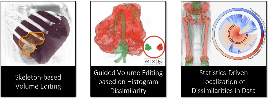
Various medical imaging techniques, such as Computed Tomography, Magnetic Resonance Imaging, Ultrasonic Imaging, are now gold standards in the diagnosis of different diseases. The diagnostic process can be greatly improved with the aid of automatic and interactive analysis tools, which, however, require certain prerequisites in order to operate. Such analysis tools can, for example, be used for pathology assessment, various standardized measurements, treatment and operation planning. One of the major requirements of such tools is the segmentation mask of an object-of-interest. However, the segmentation of medical data remains subject to errors and mistakes. Often, physicians have to manually inspect and correct the segmentation results, as (semi-)automatic techniques do not immediately satisfy the required quality. To this end, interactive segmentation editing is an integral part of medical image processing and visualization.In this thesis, we present three advanced segmentation-editing techniques. They are focused on simple interaction operations that allow the user to edit segmentation masks quickly and effectively. These operations are based on a topology-aware representation that captures structural features of the segmentation mask of the object-of-interest.
Firstly, in order to streamline the correction process, we classify segmentation defects according to underlying structural features and propose a correction procedure for each type of defect. This alleviates users from manually applying the proper editing operations, but the segmentation defects still have to be located by users.
Secondly, we extend the basic editing process by detecting regions that potentially contain defects. With subsequently suggested correction scenarios, users are hereby immediately able to correct a specific defect, instead of manually searching for defects beforehand. For each suggested correction scenario, we automatically determine the corresponding region of the respective defect in the segmentation mask and propose a suitable correction operation. In order to create the correction scenarios, we detect dissimilarities within the data values of the mask and then classify them according to the characteristics of a certain type of defect. Potential findings are presented with a glyph-based visualization that facilitates users to interactively explore the suggested correction scenarios on different levels-of-detail. As a consequence, our approach even offers users the possibility to fine-tune the chosen correction scenario instead of directly manipulating the segmentation mask, which is a time-consuming and cumbersome task.
Third and finally, we guide users through the multitude of suggested correction scenarios of the entire correction process. After statistically evaluating all suggested correction scenarios, we rank them according to their significance of dissimilarities, offering fine-grained editing capabilities at a user-specified level-of-detail. As we visually convey this ranking in a radial layout, users can easily spot and select the most (or the least) dissimilar correction scenario, which improves the segmentation mask mostly towards the desired result.
All techniques proposed within this thesis have been evaluated by collaborating radiologists. We assessed the usability, interaction aspects, the accuracy of the results and the expenditure of time of the entire correction process. The outcome of the assessment showed that our guided volume editing not only leads to acceptable segmentation results with only a few interaction steps, but also is applicable to various application scenarios.
Additional Files and Images
Weblinks
No further information available.BibTeX
@phdthesis{karimov-2016-GIVE,
  title =      "Guided Interactive Volume Editing in Medicine",
  author =     "Alexey Karimov",
  year =       "2016",
  abstract =   "Various medical imaging techniques, such as Computed
               Tomography, Magnetic Resonance Imaging, Ultrasonic Imaging,
               are now gold standards in the diagnosis of different
               diseases. The diagnostic process can be greatly improved
               with the aid of automatic and interactive analysis tools,
               which, however, require certain prerequisites in order to
               operate. Such analysis tools can, for example, be used for
               pathology assessment, various standardized measurements,
               treatment and operation planning. One of the major
               requirements of such tools is the segmentation mask of an
               object-of-interest. However, the segmentation of medical
               data remains subject to errors and mistakes. Often,
               physicians have to manually inspect and correct the
               segmentation results, as (semi-)automatic techniques do not
               immediately satisfy the required quality. To this end,
               interactive segmentation editing is an integral part of
               medical image processing and visualization.  In this thesis,
               we present three advanced segmentation-editing techniques.
               They are focused on simple interaction operations that allow
               the user to edit segmentation masks quickly and effectively.
               These operations are based on a topology-aware
               representation that captures structural features of the
               segmentation mask of the object-of-interest.  Firstly, in
               order to streamline the correction process, we classify
               segmentation defects according to underlying structural
               features and propose a correction procedure for each type of
               defect. This alleviates users from manually applying the
               proper editing operations, but the segmentation defects
               still have to be located by users.  Secondly, we extend the
               basic editing process by detecting regions that potentially
               contain defects. With subsequently suggested correction
               scenarios, users are hereby immediately able to correct a
               specific defect, instead of manually searching for defects
               beforehand. For each suggested correction scenario, we
               automatically determine the corresponding region of the
               respective defect in the segmentation mask and propose a
               suitable correction operation. In order to create the
               correction scenarios, we detect dissimilarities within the
               data values of the mask and then classify them according to
               the characteristics of a certain type of defect. Potential
               findings are presented with a glyph-based visualization that
               facilitates users to interactively explore the suggested
               correction scenarios on different levels-of-detail. As a
               consequence, our approach even offers users the possibility
               to fine-tune the chosen correction scenario instead of
               directly manipulating the segmentation mask, which is a
               time-consuming and cumbersome task.  Third and finally, we
               guide users through the multitude of suggested correction
               scenarios of the entire correction process. After
               statistically evaluating all suggested correction scenarios,
               we rank them according to their significance of
               dissimilarities, offering fine-grained editing capabilities
               at a user-specified level-of-detail. As we visually convey
               this ranking in a radial layout, users can easily spot and
               select the most (or the least) dissimilar correction
               scenario, which improves the segmentation mask mostly
               towards the desired result.  All techniques proposed within
               this thesis have been evaluated by collaborating
               radiologists. We assessed the usability, interaction
               aspects, the accuracy of the results and the expenditure of
               time of the entire correction process. The outcome of the
               assessment showed that our guided volume editing not only
               leads to acceptable segmentation results with only a few
               interaction steps, but also is applicable to various
               application scenarios.",
  month =      jun,
  address =    "Favoritenstrasse 9-11/E193-02, A-1040 Vienna, Austria",
  school =     "Institute of Computer Graphics and Algorithms, Vienna
               University of Technology ",
  URL =        "https://www.cg.tuwien.ac.at/research/publications/2016/karimov-2016-GIVE/",
}