

Haichao Miao, Gabriel Mistelbauer, Christian Nasel, Eduard Gröller
CoWRadar: Visual Quantification of the Circle of Willis in Stroke Patients
In EG Workshop on Visual Computing for Biology and Medicine, pages 1-10. September 2015.
[ demo] [
 paper]
Information
- Publication Type: Conference Paper
- Workgroup(s)/Project(s):
- Date: September 2015
- ISBN: 978-3-905674-82-8
- Publisher: The Eurographics Association
- Organization: EG Digital Library
- Location: Chester, United Kingdom
- Lecturer: Haichao Miao
- ISSN: 2070-5786
- Editor: Katja Bühler and Lars Linsen and Nigel W. John
- Booktitle: EG Workshop on Visual Computing for Biology and Medicine
- Conference date: 14. September 2015 – 15. September 2015
- Pages: 1 – 10
Abstract
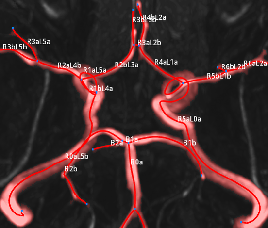
This paper presents a method for the visual quantification of cerebral arteries, known as the Circle of Willis (CoW). The CoW is an arterial structure that is responsible for the brain’s blood supply. Dysfunctions of this arterial circle can lead to strokes. The diagnosis relies on the radiologist’s expertise and the software tools used. These tools consist of very basic display methods of the volumetric data without support of advanced technologies in medical image processing and visualization. The goal of this paper is to create an automated method for the standardized description of cerebral arteries in stroke patients in order to provide an overview of the CoW’s configuration. This novel display provides visual indications of problematic areas as well as straightforward comparisons between multiple patients. Additionally, we offer a pipeline for extracting the CoW from Time-of-Flight Magnetic Resonance Angiography (TOF-MRA) data sets. An enumeration technique for the labeling of the arterial segments is therefore suggested. We also propose a method for detecting the CoW’s main supplying arteries by analyzing the coronal, sagittal and transverse image planes of the data sets. We evaluated the feasibility of our visual quantification approach in a study of 63 TOF-MRA data sets and compared our findings to those of three radiologists. The obtained results demonstrate that our proposed techniques are effective in detecting the arteries of the CoW.Additional Files and Images

Weblinks
No further information available.BibTeX
@inproceedings{Miao_2015_VCBM,
  title =      "CoWRadar: Visual Quantification of the Circle of Willis in
               Stroke Patients",
  author =     "Haichao Miao and Gabriel Mistelbauer and Christian Nasel and
               Eduard Gr\"{o}ller",
  year =       "2015",
  abstract =   "This paper presents a method for the visual quantification
               of cerebral arteries, known as the Circle of Willis (CoW).
               The CoW is an arterial structure that is responsible for the
               brain’s blood supply. Dysfunctions of this arterial circle
               can lead to strokes. The diagnosis relies on the
               radiologist’s expertise and the software tools used. These
               tools consist of very basic display methods of the
               volumetric data without support of advanced technologies in
               medical image processing and visualization. The goal of this
               paper is to create an automated method for the standardized
               description of cerebral arteries in stroke patients in order
               to provide an overview of the CoW’s configuration. This
               novel display provides visual indications of problematic
               areas as well as straightforward comparisons between
               multiple patients. Additionally, we offer a pipeline for
               extracting the CoW from Time-of-Flight Magnetic Resonance
               Angiography (TOF-MRA) data sets. An enumeration technique
               for the labeling of the arterial segments is therefore
               suggested. We also propose a method for detecting the
               CoW’s main supplying arteries by analyzing the coronal,
               sagittal and transverse image planes of the data sets. We
               evaluated the feasibility of our visual quantification
               approach in a study of 63 TOF-MRA data sets and compared our
               findings to those of three radiologists. The obtained
               results demonstrate that our proposed techniques are
               effective in detecting the arteries of the CoW.",
  month =      sep,
  isbn =       "978-3-905674-82-8",
  publisher =  "The Eurographics Association",
  organization = "EG Digital Library",
  location =   "Chester, United Kingdom",
  issn =       "2070-5786",
  editor =     "Katja B\"{u}hler and Lars Linsen and Nigel W. John",
  booktitle =  "EG Workshop on Visual Computing for Biology and Medicine",
  pages =      "1--10",
  URL =        "https://www.cg.tuwien.ac.at/research/publications/2015/Miao_2015_VCBM/",
}