

Peter Kohlmann
LiveSync: Smart Linking of 2D and 3D Views in Medical Applications
Supervisor: Eduard Gröller
Duration: March 2006 — January 2009
[image] [ screen version]
Information
- Publication Type: PhD-Thesis
- Workgroup(s)/Project(s):
- Date: 2009
- Date (Start): March 2006
- Date (End): January 2009
- TU Wien Library:
- Second Supervisor: Ivan Viola
- Rigorosum: 27. January 2009
- First Supervisor: Eduard Gröller
Abstract
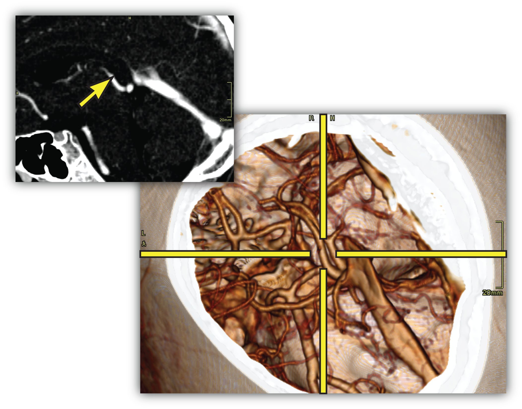
In this thesis two techniques for the smart linking of 2D and 3D views in medical applications are presented. Although real-time interactive 3D volume visualization is available even for very large data sets, it is used quite rarely in the clinical practice. A major obstacle for a better integration in the clinical workflow is the time-consuming process to adjust the parameters to generate diagnostically relevant images. The clinician has to take care of the appropriate viewpoint, zooming, transfer function setup, clipping planes, and other parameters. Because of this, current applications primarily employ 2D views generated through standard techniques such as multi-planar reformatting (MPR).The LiveSync interaction metaphor is a new concept to synchronize 2D slice views and 3D volumetric views of medical data sets. Through intuitive picking actions on the slice, the users define the anatomical structures they are interested in. The 3D volumetric view is updated automatically with the goal that the users are provided with diagnostically relevant images. To achieve this live synchronization a minimal set of derived information, without the need for segmented data sets or data-specific precomputations, is used. The presented system provides the physician with synchronized views which help to gain deeper insight into the medical data with minimal user interaction.
Contextual picking is a novel method for the interactive identification of contextual interest points within volumetric data by picking on a direct volume rendered image. In clinical diagnostics the points of interest are often located in the center of anatomical structures. In order to derive the volumetric position, which allows a convenient examination of the intended structure, the system automatically extracts contextual meta information from the DICOM (Digital Imaging and Communications in Medicine) images and the setup of the medical workstation. Along a viewing ray for a volumetric picking, the ray profile is analyzed to detect structures which are similar to predefined templates from a knowledge base. It is demonstrated that the obtained position in 3D can be utilized to highlight a structure in 2D slice views, to interactively calculate approximate centerlines of tubular objects, or to place labels at contextually-defined 3D positions.
Additional Files and Images


Weblinks
No further information available.BibTeX
@phdthesis{kohlmann-2009-lssl,
  title =      "LiveSync: Smart Linking of 2D and 3D Views in Medical
               Applications",
  author =     "Peter Kohlmann",
  year =       "2009",
  abstract =   "In this thesis two techniques for the smart linking of 2D
               and 3D views in medical applications are presented. Although
               real-time interactive 3D volume visualization is available
               even for very large data sets, it is used quite rarely in
               the clinical practice. A major obstacle for a better
               integration in the clinical workflow is the time-consuming
               process to adjust the parameters to generate diagnostically
               relevant images. The clinician has to take care of the
               appropriate viewpoint, zooming, transfer function setup,
               clipping planes, and other parameters. Because of this,
               current applications primarily employ 2D views generated
               through standard techniques such as multi-planar
               reformatting (MPR).  The LiveSync interaction metaphor is a
               new concept to synchronize 2D slice views and 3D volumetric
               views of medical data sets. Through intuitive picking
               actions on the slice, the users define the anatomical
               structures they are interested in. The 3D volumetric view is
               updated automatically with the goal that the users are
               provided with diagnostically relevant images. To achieve
               this live synchronization a minimal set of derived
               information, without the need for segmented data sets or
               data-specific precomputations, is used. The presented system
               provides the physician with synchronized views which help to
               gain deeper insight into the medical data with minimal user
               interaction.  Contextual picking is a novel method for the
               interactive identification of contextual interest points
               within volumetric data by picking on a direct volume
               rendered image. In clinical diagnostics the points of
               interest are often located in the center of anatomical
               structures. In order to derive the volumetric position,
               which allows a convenient examination of the intended
               structure, the system automatically extracts contextual meta
               information from the DICOM (Digital Imaging and
               Communications in Medicine) images and the setup of the
               medical workstation. Along a viewing ray for a volumetric
               picking, the ray profile is analyzed to detect structures
               which are similar to predefined templates from a knowledge
               base. It is demonstrated that the obtained position in 3D
               can be utilized to highlight a structure in 2D slice views,
               to interactively calculate approximate centerlines of
               tubular objects, or to place labels at contextually-defined
               3D positions.",
  address =    "Favoritenstrasse 9-11/E193-02, A-1040 Vienna, Austria",
  school =     "Institute of Computer Graphics and Algorithms, Vienna
               University of Technology ",
  URL =        "https://www.cg.tuwien.ac.at/research/publications/2009/kohlmann-2009-lssl/",
}