

 paper
paperInformation
- Publication Type: Master Thesis
- Workgroup(s)/Project(s):
- Date: March 2008
- TU Wien Library:
- First Supervisor:
- Sebastian Zambal
- Katja Bühler

- Eduard Gröller
Abstract
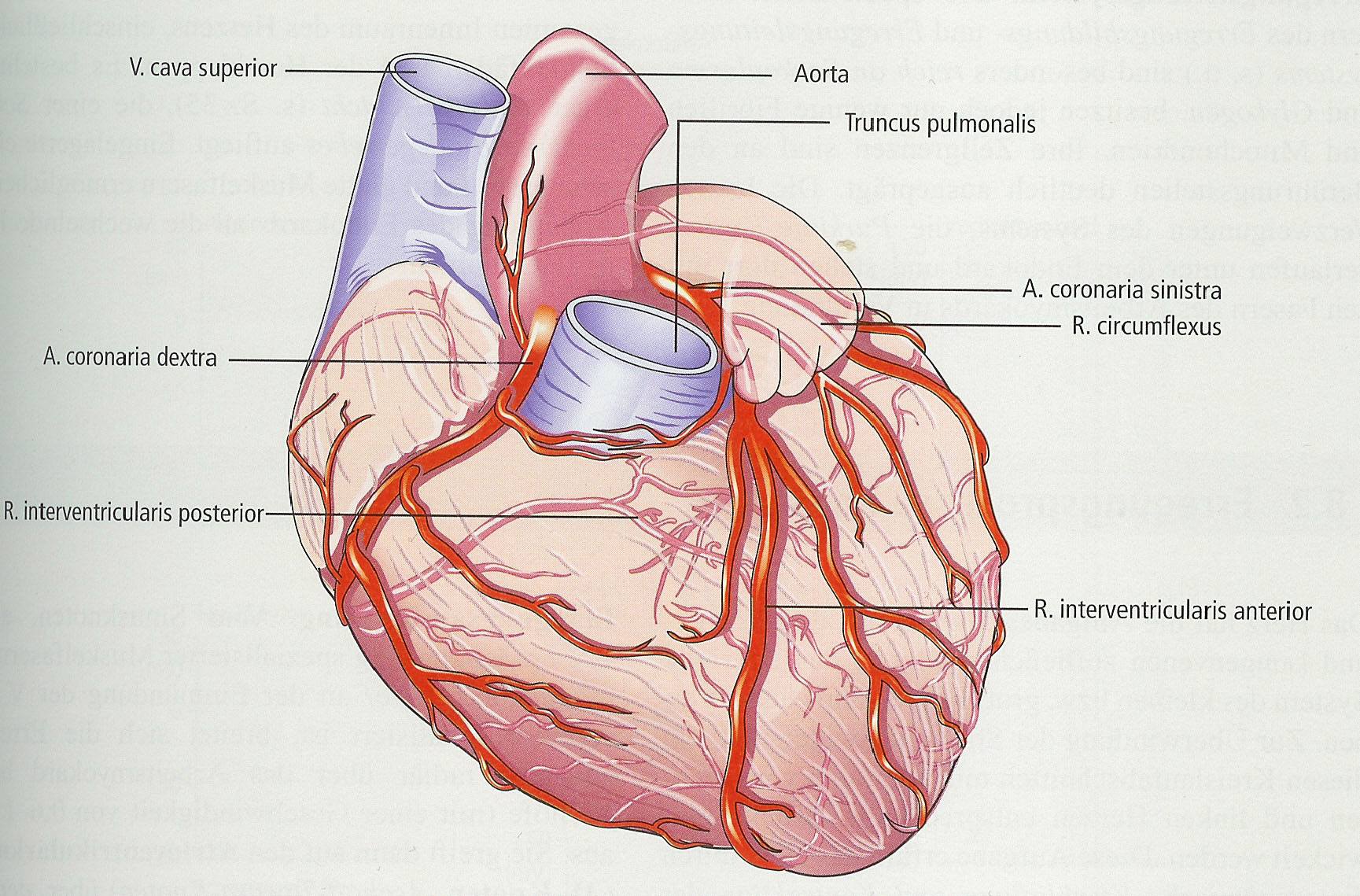
Cardiovascular diseases are the major cause of death in the developed world. About half of these are due to ischemia heart diseases. The high death rate caused by coronary artery diseases increases the need for preliminary detection. Perfusion magnetic resonance imaging has turned out to be very promising for this purpose. A contrast agent is injected intravenously to visualize the perfusion. Due to the extremely time-consuming manual analysis of these relatively large datasets, efforts for automatic approaches have been introduced. Most of these proposed methods focus on parts of the analysis process. The present thesis identifies four steps for an automatic analysis approach: localization of the heart, suppression of motion artifacts, segmentation of the myocardium, and perfusion analysis. This thesis presents a method covering all these subtasks in an automatic manner with no need for any user interaction. First the acquired MR images are analyzed to roughly detect the heart. A registration step compensates motion artifacts based on the breathing of the patient. The segmentation step provides the contour of the myocardium at every time step. Based on these segmentations the perfusion is quantified. This thesis gives a detailed description of the implementation. Furthermore the algorithm was tested on 11 datasets. The obtained results are presented and discussed. Inspection of the results indicates that this method is very promising for an efficient perfusion analysis.Additional Files and Images

Weblinks
No further information available.BibTeX
@mastersthesis{schoellhuber-2008-asc,
  title =      "Automatic Segmentation of Contrast Enhanced Cardiac MRI for
               Myocardial Perfusion Analysis",
  author =     "Andreas Sch\"{o}llhuber",
  year =       "2008",
  abstract =   "Cardiovascular diseases are the major cause of death in the
               developed world. About half of these are due to ischemia
               heart diseases. The high death rate caused by coronary
               artery diseases increases the need for preliminary
               detection. Perfusion magnetic resonance imaging has turned
               out to be very promising for this purpose. A contrast agent
               is injected intravenously to visualize the perfusion. Due to
               the extremely time-consuming manual analysis of these
               relatively large datasets, efforts for automatic approaches
               have been introduced. Most of these proposed methods focus
               on parts of the analysis process. The present thesis
               identifies four steps for an automatic analysis approach:
               localization of the heart, suppression of motion artifacts,
               segmentation of the myocardium, and perfusion analysis. This
               thesis presents a method covering all these subtasks in an
               automatic manner with no need for any user interaction.
               First the acquired MR images are analyzed to roughly detect
               the heart. A registration step compensates motion artifacts
               based on the breathing of the patient. The segmentation step
               provides the contour of the myocardium at every time step.
               Based on these segmentations the perfusion is quantified.
               This thesis gives a detailed description of the
               implementation. Furthermore the algorithm was tested on 11
               datasets. The obtained results are presented and discussed.
               Inspection of the results indicates that this method is very
               promising for an efficient perfusion analysis.",
  month =      mar,
  address =    "Favoritenstrasse 9-11/E193-02, A-1040 Vienna, Austria",
  school =     "Institute of Computer Graphics and Algorithms, Vienna
               University of Technology ",
  URL =        "https://www.cg.tuwien.ac.at/research/publications/2008/schoellhuber-2008-asc/",
}