

Caroline Magg, Laura Toussaint, Ludvig Paul Muren, Danny Indelicato, Renata Georgia Raidou
Visual Assessment of Growth Prediction in Brain Structures after Pediatric Radiotherapy
In Eurographics Workshop on Visual Computing for Biology and Medicine (VCBM2021)., pages 31-35. September 2021.
Information
- Publication Type: Conference Paper
- Workgroup(s)/Project(s):
- Date: September 2021
- Publisher: Eurographics Association
- Lecturer: Caroline Magg
- Event: EG VCBM 2021
- DOI: 10.2312/vcbm.20211343
- Call for Papers: Call for Paper
- Booktitle: Eurographics Workshop on Visual Computing for Biology and Medicine (VCBM2021).
- Pages: 5
- Pages: 31 – 35
Abstract
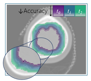
Pediatric brain tumor radiotherapy research is investigating how radiation influences the development and function of a pa- tient’s brain. To better understand how brain growth is affected by the treatment, the brain structures of the patient need to be explored and analyzed pre- and post-treatment. In this way, anatomical changes are observed over a long period, and are as- sessed as potential early markers of cognitive or functional damage. In this early work, we propose an automated approach for the visual assessment of the growth prediction of brain structures in pediatric brain tumor radiotherapy patients. Our approach reduces the need for re-segmentation, and the time required for it. We employ as a basis pre-treatment Computed Tomography (CT) scans with manual delineations (i.e., segmentation masks) of specific brain structures of interest. These pre-treatment masks are used as initialization, to predict the corresponding masks on multiple post-treatment follow-up Magnetic Resonance (MR) images, using an active contour model approach. For the accuracy quantification of the automatically predicted post- treatment masks, a support vector regressor (SVR) with features related to geometry, intensity, and gradients is trained on the pre-treatment data. Finally, a distance transform is employed to calculate the distances between pre- and post-treatment data and to visualize the predicted growth of a brain structure, along with its respective accuracy. Although segmentations of larger structures are more accurately predicted, the growth behavior of all structures is learned correctly, as indicated by the SVR results. This suggests that our pipeline is a positive initial step for the visual assessment of brain structure growth predictioAdditional Files and Images

Weblinks
BibTeX
@inproceedings{magg2021,
  title =      "Visual Assessment of Growth Prediction in Brain Structures
               after Pediatric Radiotherapy",
  author =     "Caroline Magg and Laura Toussaint and Ludvig Paul Muren and
               Danny Indelicato and Renata Georgia Raidou",
  year =       "2021",
  abstract =   "Pediatric brain tumor radiotherapy research is investigating
               how radiation influences the development and function of a
               pa- tient’s brain. To better understand how brain growth
               is affected by the treatment, the brain structures of the
               patient need to be explored and analyzed pre- and
               post-treatment. In this way, anatomical changes are observed
               over a long period, and are as- sessed as potential early
               markers of cognitive or functional damage. In this early
               work, we propose an automated approach for the visual
               assessment of the growth prediction of brain structures in
               pediatric brain tumor radiotherapy patients. Our approach
               reduces the need for re-segmentation, and the time required
               for it. We employ as a basis pre-treatment Computed
               Tomography (CT) scans with manual delineations (i.e.,
               segmentation masks) of specific brain structures of
               interest. These pre-treatment masks are used as
               initialization, to predict the corresponding masks on
               multiple post-treatment follow-up Magnetic Resonance (MR)
               images, using an active contour model approach. For the
               accuracy quantification of the automatically predicted post-
               treatment masks, a support vector regressor (SVR) with
               features related to geometry, intensity, and gradients is
               trained on the pre-treatment data. Finally, a distance
               transform is employed to calculate the distances between
               pre- and post-treatment data and to visualize the predicted
               growth of a brain structure, along with its respective
               accuracy. Although segmentations of larger structures are
               more accurately predicted, the growth behavior of all
               structures is learned correctly, as indicated by the SVR
               results. This suggests that our pipeline is a positive
               initial step for the visual assessment of brain structure
               growth predictio",
  month =      sep,
  publisher =  "Eurographics Association",
  event =      "EG VCBM 2021",
  doi =        "10.2312/vcbm.20211343",
  booktitle =  "Eurographics Workshop on Visual Computing for Biology and
               Medicine (VCBM2021).",
  pages =      "5",
  pages =      "31--35",
  URL =        "https://www.cg.tuwien.ac.at/research/publications/2021/magg2021/",
}