

 Master Thesis
Information
- Publication Type: Master Thesis
- Workgroup(s)/Project(s):
- Date: March 2019
- Date (Start): 2018
- Date (End): 5. March 2019
- TU Wien Library:
- First Supervisor: Ivan Viola

Abstract
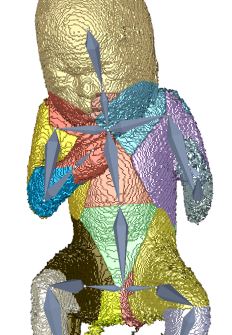
Three dimensional ultrasound images are commonly used in prenatal screening. The acquisition delivers detailed information about the skin as well as the inner organs of the fetus. Prenatal screenings in terms of growth analysis are very important to support a healthy development of the fetus. The analysis of this data involves viewing of two dimensional (2D) slices in order to take measurements or calculate the volume and weight of the fetus. These steps involve manual investigation and are dependent on the skills of the person who performs them. These measurements and calculations are very important to analyze the development of the fetus and for the birth preparation. Ultrasound imaging is a˙ected by artifacts like speckles, noise and also of structures obstructing the regions of interest. These artifacts occur because the imaging technique is using sound waves and their echo to create images. 2D slices as used as basis for the measurement of the fetus therefore might not be the best solution. Analyzing the data in a three dimensional (3D) way would enable the viewer to have a better overview and to better distinguish between artifacts and the real data of the fetus. The growth of a fetus can be analysed by comparing standardized measurements like the crown foot length, the femur length or the derived head circumference as well as the abdominal circumference. Standardization is well known in many fields of medicine and is used to enable compa-rability between investigations of the same patient or between patients. Therefore we introduce a standardized way of analyzing 3D ultrasound images of fetuses. Bringing the fetus in a standardized position would enable automatized measurements by the machine and there could also be new measurements applied like the volume of specific body parts. A standardized pose would also provide possibilities to compare the re-sults of di˙erent measurements of one fetus as well as the measurements of di˙erent fetuses. The novel method consists of six steps, namely the loading of the data, the preprocessing, the rigging of the model, the weighting of the data, the actual transformation called the "Vitruvian Baby" and at the end the analysis of the result. We tried to automatize the workflow as far as possible resulting in some manual tasks and some automatic ones. The loading of the data works with standard medical image formats and the preprocessing involves some interaction in order to get rid of the ultrasound induced artifacts. Transforming data into a specific position is a complex task which might involve a manual processing steps. In the method presented in this work one step of the transformation namely the rigging of the model, where a skeleton is placed in the data, is performed manually. The weighting as well as the transformation although are performed completely automatically resulting in a T-pose representation of the data. We analysed the performance of our novel approach in several ways. We first use a phantom model which has been used as a reference already presented in a T-pose. After using seven di˙erent fetus poses of the model as input the result was an average of 79,02%voxel overlapping between the output of the method and the goal T-pose. When having a look at the similarity of the finger to finger span and the head to toe measurement we considered a value of 91,08% and 94,05% in average. The time needed for the most complex manual task was in average seven minutes. After using a phantom model of a man, we also assessed the performance of the method using a computer model of a fetus and a phantom model of a 3D ultrasound investigation. The results also look very promising.Additional Files and Images

Weblinks
BibTeX
@mastersthesis{moerth-2018-tpose,
  title =      "Interactive Reformation of Fetal Ultrasound Data to a
               T-Position",
  author =     "Eric M\"{o}rth",
  year =       "2019",
  abstract =   "Three dimensional ultrasound images are commonly used in
               prenatal screening. The acquisition delivers detailed
               information about the skin as well as the inner organs of
               the fetus. Prenatal screenings in terms of growth analysis
               are very important to support a healthy development of the
               fetus. The analysis of this data involves viewing of two
               dimensional (2D) slices in order to take measurements or
               calculate the volume and weight of the fetus. These steps
               involve manual investigation and are dependent on the skills
               of the person who performs them. These measurements and
               calculations are very important to analyze the development
               of the fetus and for the birth preparation. Ultrasound
               imaging is a˙ected by artifacts like speckles, noise and
               also of structures obstructing the regions of interest.
               These artifacts occur because the imaging technique is using
               sound waves and their echo to create images. 2D slices as
               used as basis for the measurement of the fetus therefore
               might not be the best solution. Analyzing the data in a
               three dimensional (3D) way would enable the viewer to have a
               better overview and to better distinguish between artifacts
               and the real data of the fetus. The growth of a fetus can be
               analysed by comparing standardized measurements like the
               crown foot length, the femur length or the derived head
               circumference as well as the abdominal circumference.
               Standardization is well known in many fields of medicine and
               is used to enable compa-rability between investigations of
               the same patient or between patients. Therefore we introduce
               a standardized way of analyzing 3D ultrasound images of
               fetuses. Bringing the fetus in a standardized position would
               enable automatized measurements by the machine and there
               could also be new measurements applied like the volume of
               specific body parts. A standardized pose would also provide
               possibilities to compare the re-sults of di˙erent
               measurements of one fetus as well as the measurements of
               di˙erent fetuses. The novel method consists of six steps,
               namely the loading of the data, the preprocessing, the
               rigging of the model, the weighting of the data, the actual
               transformation called the "Vitruvian Baby" and at the end
               the analysis of the result. We tried to automatize the
               workflow as far as possible resulting in some manual tasks
               and some automatic ones. The loading of the data works with
               standard medical image formats and the preprocessing
               involves some interaction in order to get rid of the
               ultrasound induced artifacts. Transforming data into a
               specific position is a complex task which might involve a
               manual processing steps. In the method presented in this
               work one step of the transformation namely the rigging of
               the model, where a skeleton is placed in the data, is
               performed manually. The weighting as well as the
               transformation although are performed completely
               automatically resulting in a T-pose representation of the
               data. We analysed the performance of our novel approach in
               several ways. We first use a phantom model which has been
               used as a reference already presented in a T-pose. After
               using seven di˙erent fetus poses of the model as input the
               result was an average of 79,02%voxel overlapping between the
               output of the method and the goal T-pose. When having a look
               at the similarity of the finger to finger span and the head
               to toe measurement we considered a value of 91,08% and
               94,05% in average. The time needed for the most complex
               manual task was in average seven minutes. After using a
               phantom model of a man, we also assessed the performance of
               the method using a computer model of a fetus and a phantom
               model of a 3D ultrasound investigation. The results also
               look very promising.",
  month =      mar,
  address =    "Favoritenstrasse 9-11/E193-02, A-1040 Vienna, Austria",
  school =     "Research Unit of Computer Graphics, Institute of Visual
               Computing and Human-Centered Technology, Faculty of
               Informatics, TU Wien",
  URL =        "https://www.cg.tuwien.ac.at/research/publications/2019/moerth-2018-tpose/",
}