

David Bauer
Automated Visual Assessment of Osteoarthritis
[image]
Information
- Publication Type: Bachelor Thesis
- Workgroup(s)/Project(s):
- Date: July 2018
- Date (Start): 10. March 2018
- Date (End): 17. July 2018
- Matrikelnummer: 01442385
- First Supervisor: Eduard Gröller

Abstract
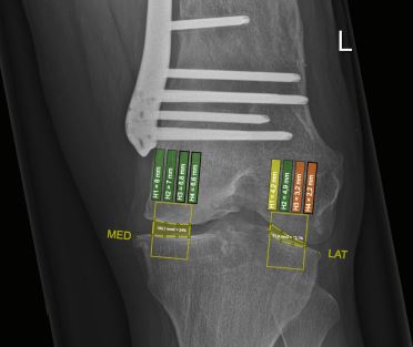
Computer-aided visualisations are a powerful tool to make large datasets more accessible. Artificial intelligence (AI) also offers diverse ways in which to extract semantic values from large data stocks. It enables users to analyse records in ways that often exceed conventional methods in their specificity and accuracy. Medicine - more specifically those specialisations requiring imaging methods - are in need of sophisticated visualisation techniques. Our team at ImageBiopsy Lab [Lju17] runs development and research in the field of AI aided visualisations in medicine. For my thesis I developed a system for measuring the joint space in x-rays of the knee, based on existing concepts. Results of the measurements are processed and presented to the user as an augmented picture. This is achieved by employing different layers of graphical overlays on top of the original image. All measurements are based on parameters of the Kellgren and Lawrence System (KLS) for classification of Osteoarthritis (OA). The proposed method enables its users to asses the stage and tendency of OA in the knee at first glance as compared to conventional methods, which can be tedious and time-consuming. Calculated focus points in the mask layers can also be adjusted in real time to accommodate for statistical outliers. The system was incorporated into an existing web-based framework which already demonstrates its potential in a clinical environment.Additional Files and Images

Weblinks
No further information available.BibTeX
@bachelorsthesis{Bauer_David_2018,
  title =      "Automated Visual Assessment of Osteoarthritis",
  author =     "David Bauer",
  year =       "2018",
  abstract =   "Computer-aided visualisations are a powerful tool to make
               large datasets more accessible. Artificial intelligence (AI)
               also offers diverse ways in which to extract semantic values
               from large data stocks. It enables users to analyse records
               in ways that often exceed conventional methods in their
               specificity and accuracy. Medicine - more specifically those
               specialisations requiring imaging methods - are in need of
               sophisticated visualisation techniques. Our team at
               ImageBiopsy Lab [Lju17] runs development and research in the
               field of AI aided visualisations in medicine. For my thesis
               I developed a system for measuring the joint space in x-rays
               of the knee, based on existing concepts. Results of the
               measurements are processed and presented to the user as an
               augmented picture. This is achieved by employing different
               layers of graphical overlays on top of the original image.
               All measurements are based on parameters of the Kellgren and
               Lawrence System (KLS) for classification of Osteoarthritis
               (OA). The proposed method enables its users to asses the
               stage and tendency of OA in the knee at first glance as
               compared to conventional methods, which can be tedious and
               time-consuming. Calculated focus points in the mask layers
               can also be adjusted in real time to accommodate for
               statistical outliers. The system was incorporated into an
               existing web-based framework which already demonstrates its
               potential in a clinical environment.",
  month =      jul,
  address =    "Favoritenstrasse 9-11/E193-02, A-1040 Vienna, Austria",
  school =     "Institute of Computer Graphics and Algorithms, Vienna
               University of Technology ",
  URL =        "https://www.cg.tuwien.ac.at/research/publications/2018/Bauer_David_2018/",
}