Information
- Publication Type: Bachelor Thesis
- Workgroup(s)/Project(s):
- Date: March 2017
- Date (Start): 1. October 2016
- Date (End): 17. March 2017
- Matrikelnummer: 1326108
- First Supervisor: Eduard Gröller
Abstract
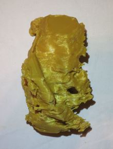
3D printing has been used industrially for decades. It enables rapid prototyping while maintaining low costs. Personal 3D printing became popular approximately since 2011. Since the massive arise of public interest, 3D printers are getting more and more affordable. In this thesis, we show how fetal 3D ultrasound data can be processed to enable 3D printing. Major steps are classification of the tissues, extraction of the isosurface and mesh-smoothing. Our approach uses thresholding, in combination with Connected Component Analysis, to separate the mother tissues from the fetal tissues. From the labeled data, we extract the fetal surface using Marching Tetrahedra. The mesh is then smoothed and converted into a data format suitable for 3D printing. Depending on the quality of the given ultrasound data, we can generate a model with recognizable facial features and peripheral structures.Additional Files and Images
Weblinks
No further information available.BibTeX
@bachelorsthesis{Wagner_032017,
  title =      "3D-Printing of Fetal Ultrasound",
  author =     "Julian Wagner",
  year =       "2017",
  abstract =   "3D printing has been used industrially for decades. It
               enables rapid prototyping while maintaining low costs.
               Personal 3D printing became popular approximately since
               2011. Since the massive arise of public interest, 3D
               printers are getting more and more affordable. In this
               thesis, we show how fetal 3D ultrasound data can be
               processed to enable 3D printing. Major steps are
               classification of the tissues, extraction of the isosurface
               and mesh-smoothing. Our approach uses thresholding, in
               combination with Connected Component Analysis, to separate
               the mother tissues from the fetal tissues. From the labeled
               data, we extract the fetal surface using Marching
               Tetrahedra. The mesh is then smoothed and converted into a
               data format suitable for 3D printing. Depending on the
               quality of the given ultrasound data, we can generate a
               model with recognizable facial features and peripheral
               structures.",
  month =      mar,
  address =    "Favoritenstrasse 9-11/E193-02, A-1040 Vienna, Austria",
  school =     "Institute of Computer Graphics and Algorithms, Vienna
               University of Technology ",
  URL =        "https://www.cg.tuwien.ac.at/research/publications/2017/Wagner_032017/",
}

 Bachelor Thesis
 image
image

