

 master thesis
Information
- Publication Type: Master Thesis
- Workgroup(s)/Project(s):
- Date: August 2014
- Date (Start): 26. August 2013
- Date (End): 26. August 2014
- TU Wien Library:
- First Supervisor: Eduard Gröller

Abstract
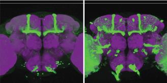
Biologists at the Institute of Molecular Pathology (IMP) in Vienna scan brains of the species Drosophila melanogaster with a confocal microscope to find relations between genes, brain structure and behavior. The database contains now more than 40.000 volumetric images, which makes it time-consuming to search for an image of interest. For biologists it would be very help-ful to have a method which can be used to search for specific images and works on the perceptual level of content. The aim of this thesis is to develop a Content Based Image Retrieval (CBIR) method customized for 3D fly brain images. A biologist can choose an image which shows interesting gene expressions and as result images which are visually similar should be retrieved. Exhaustive lit- erature research shows that in the biological field nothing comparable exists. However, CBIR plays an important role in the medical domain, which deals also with 3D images and therefore publications in this area can be seen as related. The voxelwise comparison of two images would be on the one hand computationally expensive and on the other hand not practicable due to image registration errors and anatomical variations of neuronal structures. Creating maximum intensity projections from three directions and applying a principal component analysis on the gray values overcomes the before mentioned drawbacks and delivers satisfying results. The fly brain can be divided into regions, so-called neuropils. The proposed method works on the basis of neuropils. This has, among others, the advantage that not only a global similarity can be computed, but also a comparison of images based on only some of the neuropils is possible. An extensive evaluation of the developed method is given including a parameter space exploration. For example, different lengths of the feature vectors, which describe a fly brain in a lower dimensional space, are tried and also different distance measures are tested. The evaluation shows satisfying results and that the method facilitates the work of biologists when they are looking for similar images to create a hypothesis about the connection of genes and behavior.Additional Files and Images

Weblinks
No further information available.BibTeX
@mastersthesis{Langer_Edith_IR1,
  title =      "Image Retrieval on Co-registered Confocal Microscopy Image
               Collections",
  author =     "Edith Langer",
  year =       "2014",
  abstract =   "Biologists at the Institute of Molecular Pathology (IMP) in
               Vienna scan brains of the species Drosophila  melanogaster
               with  a  confocal  microscope  to  find  relations  between
               genes,  brain structure and behavior. The database contains
               now more than 40.000 volumetric images, which makes it
               time-consuming to search for an image of interest. For
               biologists it would be very help-ful to have a method which
               can be used to search for specific images and works on the
               perceptual level of content. The aim of this thesis is to
               develop a Content Based Image Retrieval (CBIR) method
               customized for 3D fly brain images. A biologist can choose
               an image which shows interesting gene expressions and as
               result images which are visually similar should be
               retrieved.  Exhaustive lit- erature research shows that in
               the biological field nothing comparable exists.  However,
               CBIR plays an important role in the medical domain, which
               deals also with 3D images and therefore publications in this
               area can be seen as related. The voxelwise comparison of two
               images would be on the one hand computationally expensive
               and on the other hand not practicable due to image
               registration errors and anatomical variations of neuronal
               structures. Creating maximum intensity projections from
               three directions and applying a principal component analysis
               on the gray values overcomes the before mentioned drawbacks
               and delivers satisfying results. The fly brain can be
               divided into regions, so-called neuropils.  The proposed
               method works on the basis of neuropils. This has, among
               others, the advantage that not only a global similarity can
               be  computed,  but  also  a  comparison  of  images  based
               on  only  some  of  the  neuropils  is possible. An
               extensive evaluation of the developed method is given
               including a parameter space exploration.  For example,
               different lengths of the feature vectors, which describe a
               fly brain in a lower dimensional space, are tried and also
               different distance measures are tested.  The evaluation
               shows satisfying results and that the method facilitates the
               work of biologists when they are looking for similar images
               to create a hypothesis about the connection of genes and
               behavior. ",
  month =      aug,
  address =    "Favoritenstrasse 9-11/E193-02, A-1040 Vienna, Austria",
  school =     "Institute of Computer Graphics and Algorithms, Vienna
               University of Technology ",
  URL =        "https://www.cg.tuwien.ac.at/research/publications/2014/Langer_Edith_IR1/",
}