

Maurice Termeer, Javier Oliván Bescós, Marcel Breeuwer, Anna Vilanova i Bartroli , Frans Gerritsen, Eduard Gröller
The Volumetric Bulls Eye Plot
Poster shown at SCMR 2008
( 8. February 2008-10. February 2008)
In Abstracts of the 11th Annual SCMR Scientific Sessions – 2008
, pages 199-200.
[ abstract] [
 poster]
Information
- Publication Type: Poster
- Workgroup(s)/Project(s):
- Date: February 2008
- Journal: Journal of Cardiovascular Magnetic Resonance
- Volume: 11
- Series: 1
- Location: Los Angeles, California
- Event: SCMR 2008
- Booktitle: Abstracts of the 11th Annual SCMR Scientific Sessions – 2008
- Conference date: 8. February 2008 – 10. February 2008
- Pages: 199 – 200
- Keywords: Viability, Bulls Eye Plot, Cardiac MRI
Abstract
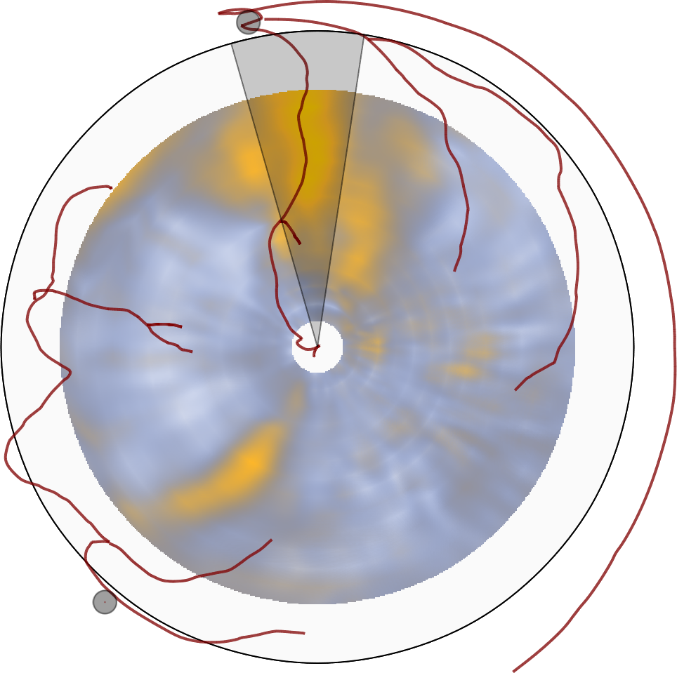
Introduction: The bull's eye plot is a commonly used schematic for the visualization of quantitative late enhancement cardiac MRI data. It gives an intuitive overview of the viability of the entire left ventricular myocardium in a single diagram. However, common implementations do not provide a continuous transition between slices and provide poor or no information about the exact location and transmurality of non-viable tissue. Purpose: We propose a novel visualization technique that relieves the drawbacks of the bull's eye plot but maintains its advantages. Our hypothesis is that our technique will enable a more accurate assessment of the relation between viable and non-viable myocardial tissue (scar). Methods: Short-axis late enhancement cardiac MRI acquisitions consist of 10-20 slices. We segment the left-ventricular myocardium in all slices using manually drawn contours on both the epicardium and the endocardium. The segmented myocardium is subsequently unfolded along the long axis and reformatted to form a thin cylinder (Figure 1a). In this process myocardial cross-sections are mapped to equidistant rings within this cylinder. The volumetric nature of the myocardium is preserved during the unfolding. A projection of the cylinder is generated using the technique of volume rendering (Figure 1b). The viewing direction in this projection is oriented from the apex towards the base of the ventricle. This makes the viewer perceive the endocardium to be behind the epicardium. This view is further augmented with the main coronary arteries extracted from a whole heart MRI scan (150 slices, SSFP). Furthermore, two dots indicating the points where the left and right-ventricular myocardial connect are added. A thin slab perpendicular to the long axis within the cylinder can be selected for exclusive rendering, providing a method of visualizing only epicardial or endocardial viability. To investigate scar transmurality, the user can select a wedge-shaped region of interest. Figure 1c shows the transmurality of that region by projecting it from its side. The unfolding method is modified for this projection to compensate for distortions due to the shape of the selected region. Since the wall thickness may vary within the region of interest, lines indicating the minimum and maximum wall thickness in the selected region are displayed. Results: The long-axis projection provides a smooth overview of the viability due to the unfolding method that preserves the continuous, volumetric nature of the myocardium. This also causes the resolution of the diagram to increase when more slices are acquired. The additional context information (i.e., coronary arteries) allows for easier interpretation of the location of any scar. Due to the close relation to the bull's eye plot, we believe that clinical adoption will be easy. The transmurality view provides detailed information on the distribution of scar within the myocardium. The preservation of wall thickness allows for judgment of the location and extent of the scar in relation to healthy tissue. Conclusion: Our novel volumetric bull's eye plot allows for a comprehensive assessment of viability and scar transmurality thanks to its continuous nature and the additional context information provided.Additional Files and Images


Weblinks
No further information available.BibTeX
@misc{termeer-2008-scmr,
  title =      "The Volumetric Bulls Eye Plot",
  author =     "Maurice Termeer and Javier Oliv\'{a}n Besc\'{o}s and Marcel
               Breeuwer and Anna Vilanova i Bartroli and Frans Gerritsen
               and Eduard Gr\"{o}ller",
  year =       "2008",
  abstract =   "Introduction: The bull's eye plot is a commonly used
               schematic for the visualization of quantitative late
               enhancement cardiac MRI data. It gives an intuitive overview
               of the viability of the entire left ventricular myocardium
               in a single diagram. However, common implementations do not
               provide a continuous transition between slices and provide
               poor or no information about the exact location and
               transmurality of non-viable tissue. Purpose: We propose a
               novel visualization technique that relieves the drawbacks of
               the bull's eye plot but maintains its advantages. Our
               hypothesis is that our technique will enable a more accurate
               assessment of the relation between viable and non-viable
               myocardial tissue (scar). Methods: Short-axis late
               enhancement cardiac MRI acquisitions consist of 10-20
               slices. We segment the left-ventricular myocardium in all
               slices using manually drawn contours on both the epicardium
               and the endocardium. The segmented myocardium is
               subsequently unfolded along the long axis and reformatted to
               form a thin cylinder (Figure 1a). In this process myocardial
               cross-sections are mapped to equidistant rings within this
               cylinder. The volumetric nature of the myocardium is
               preserved during the unfolding. A projection of the cylinder
               is generated using the technique of volume rendering (Figure
               1b). The viewing direction in this projection is oriented
               from the apex towards the base of the ventricle. This makes
               the viewer perceive the endocardium to be behind the
               epicardium. This view is further augmented with the main
               coronary arteries extracted from a whole heart MRI scan (150
               slices, SSFP). Furthermore, two dots indicating the points
               where the left and right-ventricular myocardial connect are
               added. A thin slab perpendicular to the long axis within the
               cylinder can be selected for exclusive rendering, providing
               a method of visualizing only epicardial or endocardial
               viability. To investigate scar transmurality, the user can
               select a wedge-shaped region of interest. Figure 1c shows
               the transmurality of that region by projecting it from its
               side. The unfolding method is modified for this projection
               to compensate for distortions due to the shape of the
               selected region. Since the wall thickness may vary within
               the region of interest, lines indicating the minimum and
               maximum wall thickness in the selected region are displayed.
               Results: The long-axis projection provides a smooth overview
               of the viability due to the unfolding method that preserves
               the continuous, volumetric nature of the myocardium. This
               also causes the resolution of the diagram to increase when
               more slices are acquired. The additional context information
               (i.e., coronary arteries) allows for easier interpretation
               of the location of any scar. Due to the close relation to
               the bull's eye plot, we believe that clinical adoption will
               be easy. The transmurality view provides detailed
               information on the distribution of scar within the
               myocardium. The preservation of wall thickness allows for
               judgment of the location and extent of the scar in relation
               to healthy tissue. Conclusion: Our novel volumetric bull's
               eye plot allows for a comprehensive assessment of viability
               and scar transmurality thanks to its continuous nature and
               the additional context information provided. ",
  month =      feb,
  journal =    "Journal of Cardiovascular Magnetic Resonance",
  volume =     "11",
  series =     "1",
  location =   "Los Angeles, California",
  event =      "SCMR 2008",
  booktitle =  "Abstracts of the 11th Annual SCMR Scientific Sessions –
               2008",
  Conference date = "Poster presented at SCMR 2008 (2008-02-08--2008-02-10)",
  note =       "199--200",
  pages =      "199 – 200",
  keywords =   "Viability, Bulls Eye Plot, Cardiac MRI",
  URL =        "https://www.cg.tuwien.ac.at/research/publications/2008/termeer-2008-scmr/",
}