

Armin Kanitsar, Dominik Fleischmann, Rainer Wegenkittl, Eduard Gröller
Diagnostic Relevant Visualization of Vascular Structures
In Scientific Visualization: The Visual Extraction of Knowledge from Data, pages 207-228, 2005
[ PDF]
PDF]
Information
- Publication Type: Article in a Book
- Workgroup(s)/Project(s):
- Date: 2005
- Booktitle: Scientific Visualization: The Visual Extraction of Knowledge from Data
- Editor: G.-P. Bonneau, T. Ertl, G.M. Nielson
- ISBN: 3540260668
- Publisher: Springer Verlag, Berlin
- Pages: 207 – 228
Abstract
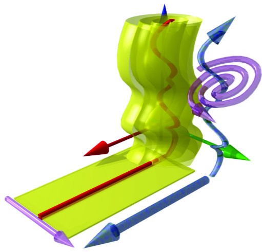
Traditional volume visualization techniques sometimes provide incomplete clinical information needed for applications in medical visualization. In the area of vascular visualization important features such as the lumen of a diseased vessel segment may not be visible. One way to display vascular structures for diagnostic purposes is to generate longitudinal cross-sections in order to show their lumen, wall, and surrounding tissue in a curved plane. Curved planar reformation (CPR) has proven to be an acceptable practical solution. We discuss four different methods to generate CPR images from single vessel segments: Projected CPR, stretched CPR, straightened CPR, and helical CPR. Furthermore we investigate three different methods for displaying vascular trees: Multi-path projected CPR, multi-path stretched CPR, and untangled CPR. The principle concept of each method is discussed and detailed information for the realization is given. In addition the properties, advantages and disadvantages of each method are summarized.Additional Files and Images

Weblinks
No further information available.BibTeX
@incollection{groeller-2005-dia,
  title =      "Diagnostic Relevant Visualization of Vascular Structures ",
  author =     "Armin Kanitsar and Dominik Fleischmann and Rainer Wegenkittl
               and Eduard Gr\"{o}ller",
  year =       "2005",
  abstract =   "Traditional volume visualization techniques sometimes
               provide incomplete clinical information needed for
               applications in medical visualization. In the area of
               vascular visualization important features such as the lumen
               of a diseased vessel segment may not be visible. One way to
               display vascular structures for diagnostic purposes is to
               generate longitudinal cross-sections in order to show their
               lumen, wall, and surrounding tissue in a curved plane.
               Curved planar reformation (CPR) has proven to be an
               acceptable practical solution. We discuss four different
               methods to generate CPR images from single vessel segments:
               Projected CPR, stretched CPR, straightened CPR, and helical
               CPR. Furthermore we investigate three different methods for
               displaying vascular trees: Multi-path projected CPR,
               multi-path stretched CPR, and untangled CPR. The principle
               concept of each method is discussed and detailed information
               for the realization is given. In addition the properties,
               advantages and disadvantages of each method are summarized.",
  booktitle =  "Scientific Visualization: The Visual Extraction of Knowledge
               from Data",
  editor =     "G.-P. Bonneau, T. Ertl, G.M. Nielson",
  isbn =       "3540260668",
  publisher =  "Springer Verlag, Berlin",
  URL =        "https://www.cg.tuwien.ac.at/research/publications/2005/groeller-2005-dia/",
}