

Thomas Tramberger
Automatic Breast Lesion Evaluation for Comparative Studies
[ Bachelor Thesis] [image]
Information
- Publication Type: Bachelor Thesis
- Workgroup(s)/Project(s):
- Date: May 2018
- Date (Start): 1. December 2017
- Date (End): 8. May 2018
- Matrikelnummer: 01326870
- First Supervisor: Renata Georgia Raidou

Abstract
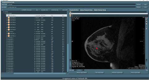
Breast cancer is the most common cancer with a high mortality rate. Neoadjuvant chemotherapie is conducted before surgery to reduce the breast tumor mass. Currently, a lot of trials are taking place, with the purpose of understanding the effects of different chemotherapy strategies. In this work a software is developed to analyse and compare the influence of these treatments. The study data is available as 4D Dynamic Contrast-Enhanced Magnetic Resonance Imaging data. To reduce the time of manual segmentation and the connection of segmented lesions over time a automatic procedure was implemented. This process uses the time-signal intensity curve and a support vector machine to classify lesions with calculated morphological features. To analyse the data, two views are available. The Intra-patient view visualizes the tumor behaviour of an individual patient over time. With the Multi-patient view the user is able to compare multiple patients’ lesions and additional added patient data. Both views are implemented with JavaScript and can be expanded easily. Because of missing ground truth an evaluation of the automatic segmentation method was not possible.Additional Files and Images

Weblinks
No further information available.BibTeX
@bachelorsthesis{Tramberger_2018,
  title =      "Automatic Breast Lesion Evaluation for Comparative Studies",
  author =     "Thomas Tramberger",
  year =       "2018",
  abstract =   "Breast cancer is the most common cancer with a high
               mortality rate. Neoadjuvant chemotherapie is conducted
               before surgery to reduce the breast tumor mass. Currently, a
               lot of trials are taking place, with the purpose of
               understanding the effects of different chemotherapy
               strategies. In this work a software is developed to analyse
               and compare the influence of these treatments. The study
               data is available as 4D Dynamic Contrast-Enhanced Magnetic
               Resonance Imaging data. To reduce the time of manual
               segmentation and the connection of segmented lesions over
               time a automatic procedure was implemented. This process
               uses the time-signal intensity curve and a support vector
               machine to classify lesions with calculated morphological
               features. To analyse the data, two views are available. The
               Intra-patient view visualizes the tumor behaviour of an
               individual patient over time. With the Multi-patient view
               the user is able to compare multiple patients’ lesions and
               additional added patient data. Both views are implemented
               with JavaScript and can be expanded easily. Because of
               missing ground truth an evaluation of the automatic
               segmentation method was not possible.",
  month =      may,
  address =    "Favoritenstrasse 9-11/E193-02, A-1040 Vienna, Austria",
  school =     "Institute of Computer Graphics and Algorithms, Vienna
               University of Technology ",
  URL =        "https://www.cg.tuwien.ac.at/research/publications/2018/Tramberger_2018/",
}