

Renata Raidou , Freek Marcelis, Marcel Breeuwer, Eduard Gröller, Anna Vilanova i Bartroli, Huub van de Wetering
Visual Analytics for the Exploration and Assessment of Segmentation Errors
Eurographics Workshop on Visual Computing for Biology and Medicine:193-202, September 2016. [image] [ Paper]
Information
- Publication Type: Journal Paper with Conference Talk
- Workgroup(s)/Project(s):
- Date: September 2016
- Journal: Eurographics Workshop on Visual Computing for Biology and Medicine
- Lecturer:
- Pages: 193 – 202
Abstract
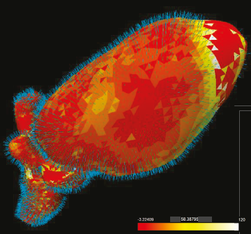
Several diagnostic and treatment procedures require the segmentation of anatomical structures from medical images. However, the automatic model-based methods that are often employed, may produce inaccurate segmentations. These, if used as input for diagnosis or treatment, can have detrimental effects for the patients. Currently, an analysis to predict which anatomic regions are more prone to inaccuracies, and to determine how to improve segmentation algorithms, cannot be performed. We propose a visual tool to enable experts, working on model-based segmentation algorithms, to explore and analyze the outcomes and errors of their methods. Our approach supports the exploration of errors in a cohort of pelvic organ segmentations, where the performance of an algorithm can be assessed. Also, it enables the detailed exploration and assessment of segmentation errors, in individual subjects. To the best of our knowledge, there is no other tool with comparable functionality. A usage scenario is employed to explore and illustrate the capabilities of our visual tool. To further assess the value of the proposed tool, we performed an evaluation with five segmentation experts. The evaluation participants confirmed the potential of the tool in providing new insight into their data and employed algorithms. They also gave feedback for future improvements.Additional Files and Images

Weblinks
No further information available.BibTeX
@article{Groeller_2016_P4,
  title =      "Visual Analytics for the Exploration and Assessment  of
               Segmentation Errors",
  author =     "Renata Raidou and Freek Marcelis and Marcel Breeuwer and
               Eduard Gr\"{o}ller and Anna Vilanova i Bartroli and Huub van
               de Wetering",
  year =       "2016",
  abstract =   "Several diagnostic and treatment procedures require the
               segmentation of anatomical structures from medical images.
               However, the automatic model-based methods that are often
               employed, may produce inaccurate segmentations. These, if
               used as input for diagnosis or treatment, can have
               detrimental effects for the patients. Currently, an analysis
               to predict which anatomic regions are more prone to
               inaccuracies, and to determine how to improve segmentation
               algorithms, cannot be performed. We propose a visual tool to
               enable experts, working on model-based segmentation
               algorithms, to explore and analyze the outcomes and errors
               of their methods. Our approach supports the exploration of
               errors in a cohort of pelvic organ segmentations, where the
               performance of an algorithm can be assessed. Also, it
               enables the detailed exploration and assessment of
               segmentation errors, in individual subjects. To the best of
               our knowledge, there is no other tool with comparable
               functionality. A usage scenario is employed to explore and
               illustrate the capabilities of our visual tool. To further
               assess the value of the proposed tool, we performed an
               evaluation with five segmentation experts. The evaluation
               participants confirmed the potential of the tool in
               providing new insight into their data and employed
               algorithms. They also gave feedback for future improvements.",
  month =      sep,
  journal =    "Eurographics Workshop on Visual Computing for Biology and
               Medicine",
  pages =      "193--202",
  URL =        "https://www.cg.tuwien.ac.at/research/publications/2016/Groeller_2016_P4/",
}