Information
- Publication Type: PhD-Thesis
- Workgroup(s)/Project(s):
- Date: October 2012
- Date (Start): September 2009
- Date (End): September 2012
- TU Wien Library:
- 1st Reviewer: Eduard Gröller
- 2nd Reviewer: Milos Sramek
- Rigorosum: 24. October 2012
- First Supervisor: Eduard Gröller
- Keywords: ultrasound, volume rendering, medical imaging
Abstract
Ultrasound (US) imaging is due to its real-time character, low cost, non-invasive nature, high availability, and many other factors, considered a standard diagnostic procedure during pregnancy. The quality of diagnostics depends on many factors, including scanning protocol, data characteristics and visualization algorithms. In this work, several problems of ultrasound data visualization for obstetric ultrasound imaging are discussed and addressed.The capability of ultrasound scanners is growing and modern ultrasound devices produce large amounts of data that have to be processed in real-time. An ultrasound imaging system is in a broad sense a pipeline of several operations and visualization algorithms. Individual algorithms are usually organized in modules that separately process the data. In order to achieve the required level of detail and high quality images with the visualization pipeline, we had to address the flow of large amounts of data on modern computer hardware with limited capacity. We developed a novel architecture of visualization pipeline for ultrasound imaging. This visualization pipeline combines several algorithms, which are described in this work, into the integrated system. In the context of this pipeline, we advocate slice-based streaming as a possible approach for the large data flow problem.
Live examination of the moving fetus from ultrasound data is a challenging task which requires extensive knowledge of the fetal anatomy and a proficient operation of the ultrasound machine. The fetus is typically occluded by structures which hamper the view in 3D rendered images. We developed a novel method of visualizing the human fetus for prenatal sonography from 3D/4D ultrasound data. It is a fully automatic method that can recognize and render the fetus without occlusion, where the highest priority is to achieve an unobstructed view of the fetal face. Our smart visibility method for prenatal ultrasound is based on a ray-analysis performed within image-based direct volume rendering (DVR). It automatically calculates a clipping surface that removes the uninteresting structures and uncovers the interesting structures of the fetal anatomy behind. The method is able to work with the data streamed on-the-fly from the ultrasound transducer and to visualize a temporal sequence of reconstructed ultrasound data in real time. It has the potential to minimize the interaction of the operator and to improve the comfort of patients by decreasing the investigation time. This can lead to an increased confidence in the prenatal diagnosis with 3D ultrasound and eventually decrease the costs of the investigation.
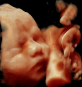
Ultrasound scanning is very popular among parents who are interested in the health condition of their fetus during pregnancy. Parents usually want to keep the ultrasound images as a memory for the future. Furthermore, convincing images are important for the confident communication of findings between clinicians and parents. Current ultrasound devices offer advanced imaging capabilities, but common visualization methods for volumetric data only provide limited visual fidelity. The standard methods render only images with a plastic-like appearance which do not correspond to naturally looking fetuses. This is partly due to the dynamic and noisy nature of the data which limits the applicability of standard volume visualization techniques. In this thesis, we present a fetoscopic rendering method which aims to reproduce the quality of fetoscopic examinations (i.e., physical endoscopy of the uterus) from 4D sonography data. Based on the requirements of domain experts and the constraints of live ultrasound imaging, we developed a method for high-quality rendering of prenatal examinations. We employ a realistic illumination model which supports shadows, movable light sources, and realistic rendering of the human skin to provide an immersive experience for physicians and parents alike. Beyond aesthetic aspects, the resulting visualizations have also promising diagnostic applications. The presented fetoscopic rendering method has been successfully integrated in the state-of-the-art ultrasound imaging systems of GE Healthcare as HDlive imaging tool. It is daily used in many prenatal imaging centers around the world.
Additional Files and Images
Weblinks
No further information available.BibTeX
@phdthesis{varchola_andrej-2012-fetoscopic,
  title =      "Live Fetoscopic Visualization of 4D Ultrasound Data",
  author =     "Andrej Varchola",
  year =       "2012",
  abstract =   "Ultrasound (US) imaging is due to its real-time character,
               low cost, non-invasive nature, high availability, and many
               other factors, considered a standard diagnostic procedure
               during pregnancy. The quality of diagnostics depends on many
               factors, including scanning protocol, data characteristics
               and visualization algorithms. In this work, several problems
               of ultrasound data visualization for obstetric ultrasound
               imaging are discussed and addressed.  The capability of
               ultrasound scanners is growing and modern ultrasound devices
               produce large amounts of data that have to be processed in
               real-time. An ultrasound imaging system is in a broad sense
               a pipeline of several operations and visualization
               algorithms. Individual algorithms are usually organized in
               modules that separately process the data. In order to
               achieve the required level of detail and high quality images
               with the visualization pipeline, we had to address the flow
               of large amounts of data on modern computer hardware with
               limited capacity. We developed a novel architecture of
               visualization pipeline for ultrasound imaging. This
               visualization pipeline combines several algorithms, which
               are described in this work, into the integrated system. In
               the context of this pipeline, we advocate slice-based
               streaming as a possible approach for the large data flow
               problem.  Live examination of the moving fetus from
               ultrasound data is a challenging task which requires
               extensive knowledge of the fetal anatomy and a proficient
               operation of the ultrasound machine. The fetus is typically
               occluded by structures which hamper the view in 3D rendered
               images. We developed a novel method of visualizing the human
               fetus for prenatal sonography from 3D/4D ultrasound data. It
               is a fully automatic method that can recognize and render
               the fetus without occlusion, where the highest priority is
               to achieve an unobstructed view of the fetal face. Our smart
               visibility method for prenatal ultrasound is based on a
               ray-analysis performed within image-based direct volume
               rendering (DVR). It automatically calculates a clipping
               surface that removes the uninteresting structures and
               uncovers the interesting structures of the fetal anatomy
               behind. The method is able to work with the data streamed
               on-the-fly from the ultrasound transducer and to visualize a
               temporal sequence of reconstructed ultrasound data in real
               time. It has the potential to minimize the interaction of
               the operator and to improve the comfort of patients by
               decreasing the investigation time. This can lead to an
               increased confidence in the prenatal diagnosis with 3D
               ultrasound and eventually decrease the costs of the
               investigation.  Ultrasound scanning is very popular among
               parents who are interested in the health condition of their
               fetus during pregnancy. Parents usually want to keep the
               ultrasound images as a memory for the future. Furthermore,
               convincing images are important for the confident
               communication of findings between clinicians and parents.
               Current ultrasound devices offer advanced imaging
               capabilities, but common visualization methods for
               volumetric data only provide limited visual fidelity. The
               standard methods render only images with a plastic-like
               appearance which do not correspond to naturally looking
               fetuses. This is partly due to the dynamic and noisy nature
               of the data which limits the applicability of standard
               volume visualization techniques. In this thesis, we present
               a fetoscopic rendering method which aims to reproduce the
               quality of fetoscopic examinations (i.e., physical endoscopy
               of the uterus) from 4D sonography data. Based on the
               requirements of domain experts and the constraints of live
               ultrasound imaging, we developed a method for high-quality
               rendering of prenatal examinations. We employ a realistic
               illumination model which supports shadows, movable light
               sources, and realistic rendering of the human skin to
               provide an immersive experience for physicians and parents
               alike. Beyond aesthetic aspects, the resulting
               visualizations have also promising diagnostic applications.
               The presented fetoscopic rendering method has been
               successfully integrated in the state-of-the-art ultrasound
               imaging systems of GE Healthcare as HDlive imaging tool. It
               is daily used in many prenatal imaging centers around the
               world.",
  month =      oct,
  address =    "Favoritenstrasse 9-11/E193-02, A-1040 Vienna, Austria",
  school =     "Institute of Computer Graphics and Algorithms, Vienna
               University of Technology ",
  keywords =   "ultrasound, volume rendering, medical imaging",
  URL =        "https://www.cg.tuwien.ac.at/research/publications/2012/varchola_andrej-2012-fetoscopic/",
}


 Thesis
