

Maurice Termeer, Javier Oliván Bescós, Marcel Breeuwer, Anna Vilanova i Bartroli , Frans Gerritsen, Eduard Gröller
CoViCAD: Comprehensive Visualization of Coronary Artery Disease
IEEE Transactions on Visualization and Computer Graphics (accepted for publication), 13(6), October 2007. [ paper]
Information
- Publication Type: Journal Paper with Conference Talk
- Workgroup(s)/Project(s):
- Date: October 2007
- Journal: IEEE Transactions on Visualization and Computer Graphics (accepted for publication)
- Volume: 13
- Number: 6
- Lecturer: Maurice Termeer
- Keywords: VBEP, viability, late enhancement, Cardiac MRI, bull
Abstract
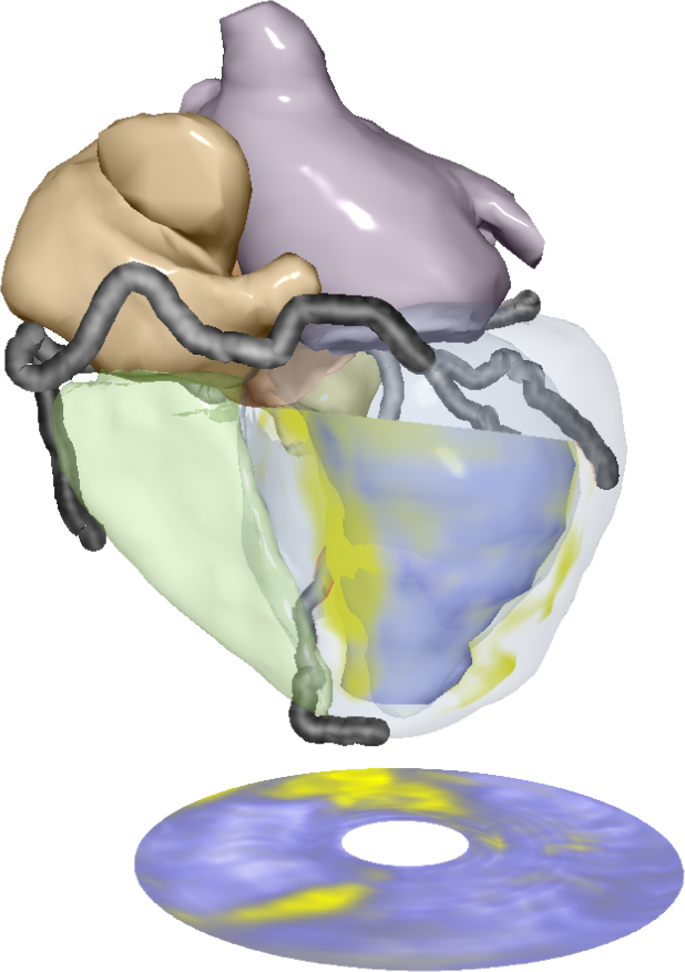
We present novel, comprehensive visualization techniques for the diagnosis of patients with Coronary Artery Disease using segmented cardiac MRI data. We extent an accepted medical visualization technique called the bull’s eye plot by removing discontinuities, preserving the volumetric nature of the left ventricular wall and adding anatomical context. The resulting volumetric bull’s eye plot can be used for the assessment of transmurality. We link these visualizations to a 3D view that presents viability information in a detailed anatomical context. We combine multiple MRI scans (whole heart anatomical data, late enhancement data) and multiple segmentations (polygonal heart model, late enhancement contours, coronary artery tree). By selectively combining different rendering techniques we obtain comprehensive yet intuitive visualizations of the various data sources.Additional Files and Images
Additional images and videos
Additional files

Weblinks
No further information available.BibTeX
@article{termeer-2007-covicad,
  title =      "CoViCAD: Comprehensive Visualization of Coronary Artery
               Disease",
  author =     "Maurice Termeer and Javier Oliv\'{a}n Besc\'{o}s and Marcel
               Breeuwer and Anna Vilanova i Bartroli and Frans Gerritsen
               and Eduard Gr\"{o}ller",
  year =       "2007",
  abstract =   "We present novel, comprehensive visualization techniques for
               the diagnosis of patients with Coronary Artery Disease using
               segmented cardiac MRI data. We extent an accepted medical
               visualization technique called the bull’s eye plot by
               removing discontinuities, preserving the volumetric nature
               of the left ventricular wall and adding anatomical context.
               The resulting volumetric bull’s eye plot can be used for
               the assessment of transmurality. We link these
               visualizations to a 3D view that presents viability
               information in a detailed anatomical context. We combine
               multiple MRI scans (whole heart anatomical data, late
               enhancement data) and multiple segmentations (polygonal
               heart model, late enhancement contours, coronary artery
               tree). By selectively combining different rendering
               techniques we obtain comprehensive yet intuitive
               visualizations of the various data sources.",
  month =      oct,
  journal =    "IEEE Transactions on Visualization and Computer Graphics
               (accepted for publication)",
  volume =     "13",
  number =     "6",
  keywords =   "VBEP, viability, late enhancement, Cardiac MRI, bull",
  URL =        "https://www.cg.tuwien.ac.at/research/publications/2007/termeer-2007-covicad/",
}