

Matús Straka
Processing and Visualization of Peripheral CT-Angiography Datasets
Supervisor: Milos Sramek
Duration: April 2002 — August 2006
[] [ pdf]
pdf]
Information
- Publication Type: PhD-Thesis
- Workgroup(s)/Project(s): not specified
- Date: August 2006
- Date (Start): April 2002
- Date (End): August 2006
- TU Wien Library:
- Second Supervisor: Eduard Gröller

- Rigorosum: 27. August 2006
- First Supervisor: Milos Sramek
- Keywords: Visualization, Medial Data Processing, Segmentation, Vessel Modeling, 3D Reconstruction, Vessel Visualization
Abstract
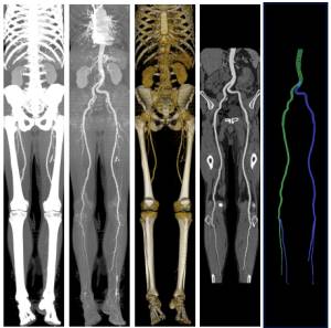
In this thesis, individual steps of a pipeline for processing of the peripheral Computed Tomography Angiography (CTA) datasets are addressed. The peripheral CTA datasets are volumetric datasets representing pathologies in vascularity of the lower extremities in the human body. These pathologies result from various atherosclerotic diseases as e.g. the Peripheral Arterial Occlusive Disease (PAOD) and their early and precise diagnostics significantly contributes to planning of a later interventional radiology treatment.The diagnostics is based on visualization of the imaged vascular tree, where individual pathologic changes, as plaque, calcifications, stenoses of the vessel lumen and occluded parts of the vessels are visible. CTA has evolved within the recent years into a robust, accurate and cost effective imaging technique for patients with both coronary and arterial diseases. As a result of the CTA scanning, a set of 1200–2000 transverse slices is acquired, depicting vessels enhanced by means of an intravenously injected contrast medium. The number of slices is high and therefore their manual examination is laborious and lengthy. As a remedy, post-processing methods were developed to allow faster and more intuitive visualization of the imaged vascularity. However, simple visualization by means of the traditional techniques as maximum-intensity projection (MIP) or direct volume rendering (DVR) is hampered due to the presence of bones in the dataset, which occlude the vessels. Therefore, a sequence of operations—the processing pipeline—is needed, leading to generation of clinically relevant images which depict unobstructed vessels.
In the first step of the pipeline the dataset is segmented and the tissues are classified, to allow subsequent vessel identification and bone removal. This is a complex task because of high density and spatial variability of the tissues. Traditional image processing techniques do not deliver acceptable results and therefore in the thesis we present new approaches that introduce additional ’anatomic’ information into the segmentation and classification process. We propose a probabilistic atlas which enables modeling of spatial and density distributions of vessel and bone tissues in datasets, to allow their improved classification. In the atlas construction the non-rigid thin-plate spline warping and registration of the datasets are applied, to address the high anatomic variability among the patients. The concept of the atlas is further extended by means of the watershed transform, to further improve precision of the registration procedure. Alternatively, we propose and evaluate a technique for vessel enhancement based on Hessian filtering to allow detection and recognition of vessel structures without operator supervision.
In the second step a geometric model of the vessel tree is constructed to derive information about the vessel centerlines. Here, an already available algorithm based on the so-called vessel-tracking, implemented by means of optimal path searching, is exploited with improvements to make the geometric model more precise.
The third step of the processing pipeline—visualization—requires this model, since its results can be significantly influenced by a potential imperfections, bringing in clinically misleading images. To address limitations of the vessel visualization by means of the existing techniques as MIP, CPR or DVR we propose their generalization in form of a focus & context-based concept called VesselGlyph. VesselGlyph enables to combine intuitively and systematically various visualization techniques to single a image to allow better, more comprehensive and unoccluded view of vessels for the diagnostic purposes.
To support the design and development of the proposed segmentation, modeling and visualization algorithms and to enable their application in the clinical environment, we implemented a set of tools grouped in the AngioVis ToolBox software. Within this application, individual steps of the processing pipeline are accomplished. The toolbox is complemented with additional utilities constituting together a fully-functional medical workstation software which is regularly used to process patient data on a daily basis in the clinical environment.
Additional Files and Images

Weblinks
No further information available.BibTeX
@phdthesis{straka-phd-thesis,
  title =      "Processing and Visualization of Peripheral CT-Angiography
               Datasets",
  author =     "Mat\'{u}s Straka",
  year =       "2006",
  abstract =   "In this thesis, individual steps of a pipeline for
               processing of the peripheral Computed Tomography Angiography
               (CTA) datasets are addressed. The peripheral CTA datasets
               are volumetric datasets representing pathologies in
               vascularity of the lower extremities in the human body.
               These pathologies result from various atherosclerotic
               diseases as e.g. the Peripheral Arterial Occlusive Disease
               (PAOD) and their early and precise diagnostics significantly
               contributes to planning of a later interventional radiology
               treatment.  The diagnostics is based on visualization of the
               imaged vascular tree, where individual pathologic changes,
               as plaque, calcifications, stenoses of the vessel lumen and
               occluded parts of the vessels are visible. CTA has evolved
               within the recent years into a robust, accurate and cost
               effective imaging technique for patients with both coronary
               and arterial diseases. As a result of the CTA scanning, a
               set of 1200–2000 transverse slices is acquired, depicting
               vessels enhanced by means of an intravenously injected
               contrast medium. The number of slices is high and therefore
               their manual examination is laborious and lengthy. As a
               remedy, post-processing methods were developed to allow
               faster and more intuitive visualization of the imaged
               vascularity. However, simple visualization by means of the
               traditional techniques as maximum-intensity projection (MIP)
               or direct volume rendering (DVR) is hampered due to the
               presence of bones in the dataset, which occlude the vessels.
               Therefore, a sequence of operations—the processing
               pipeline—is needed, leading to generation of clinically
               relevant images which depict unobstructed vessels.  In the
               first step of the pipeline the dataset is segmented and the
               tissues are classified, to allow subsequent vessel
               identification and bone removal. This is a complex task
               because of high density and spatial variability of the
               tissues. Traditional image processing techniques do not
               deliver acceptable results and therefore in the thesis we
               present new approaches that introduce additional
               ’anatomic’ information into the segmentation and
               classification process. We propose a probabilistic atlas
               which enables modeling of spatial and density distributions
               of vessel and bone tissues in datasets, to allow their
               improved classification. In the atlas construction the
               non-rigid thin-plate spline warping and registration of the
               datasets are applied, to address the high anatomic
               variability among the patients. The concept of the atlas is
               further extended by means of the watershed transform, to
               further improve precision of the registration procedure.
               Alternatively, we propose and evaluate a technique for
               vessel enhancement based on Hessian filtering to allow
               detection and recognition of vessel structures without
               operator supervision.  In the second step a geometric model
               of the vessel tree is constructed to derive information
               about the vessel centerlines. Here, an already available
               algorithm based on the so-called vessel-tracking,
               implemented by means of optimal path searching, is exploited
               with improvements to make the geometric model more precise.
               The third step of the processing
               pipeline—visualization—requires this model, since its
               results can be significantly influenced by a potential
               imperfections, bringing in clinically misleading images. To
               address limitations of the vessel visualization by means of
               the existing techniques as MIP, CPR or DVR we propose their
               generalization in form of a focus & context-based concept
               called VesselGlyph. VesselGlyph enables to combine
               intuitively and systematically various visualization
               techniques to single a image to allow better, more
               comprehensive and unoccluded view of vessels for the
               diagnostic purposes.  To support the design and development
               of the proposed segmentation, modeling and visualization
               algorithms and to enable their application in the clinical
               environment, we implemented a set of tools grouped in the
               AngioVis ToolBox software. Within this application,
               individual steps of the processing pipeline are
               accomplished. The toolbox is complemented with additional
               utilities constituting together a fully-functional medical
               workstation software which is regularly used to process
               patient data on a daily basis in the clinical environment.",
  month =      aug,
  address =    "Favoritenstrasse 9-11/E193-02, A-1040 Vienna, Austria",
  school =     "Institute of Computer Graphics and Algorithms, Vienna
               University of Technology ",
  keywords =   "Visualization, Medial Data Processing, Segmentation, Vessel
               Modeling, 3D Reconstruction, Vessel Visualization",
  URL =        "https://www.cg.tuwien.ac.at/research/publications/2006/straka-phd-thesis/",
}