Data Acquisition
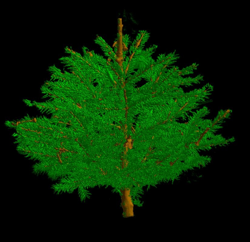
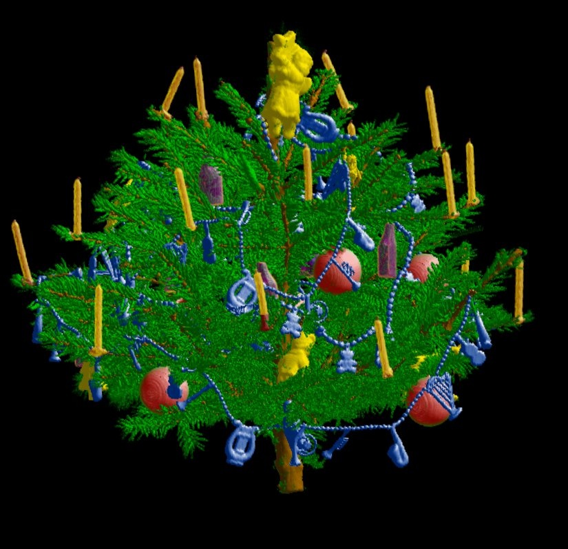
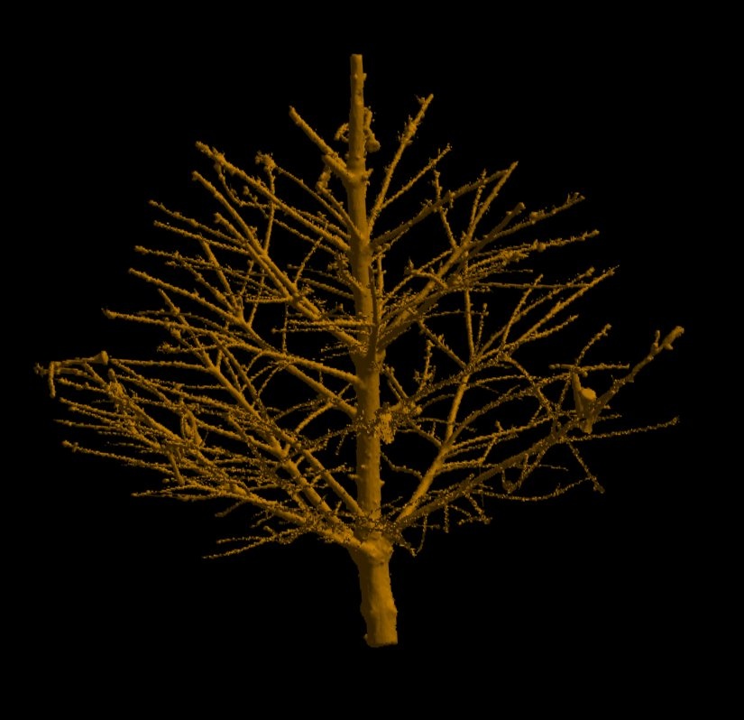
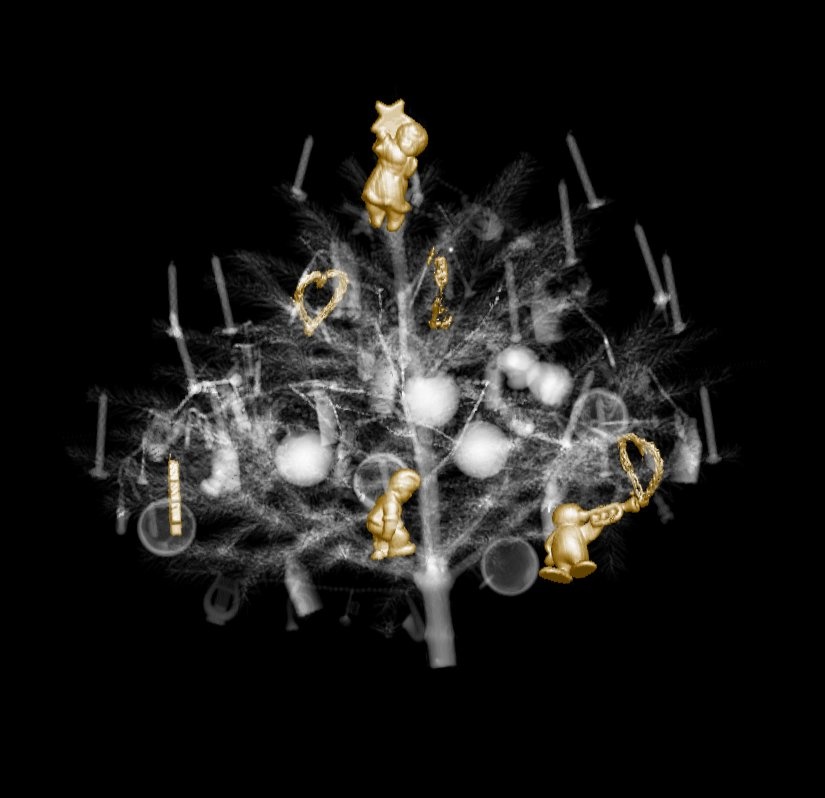
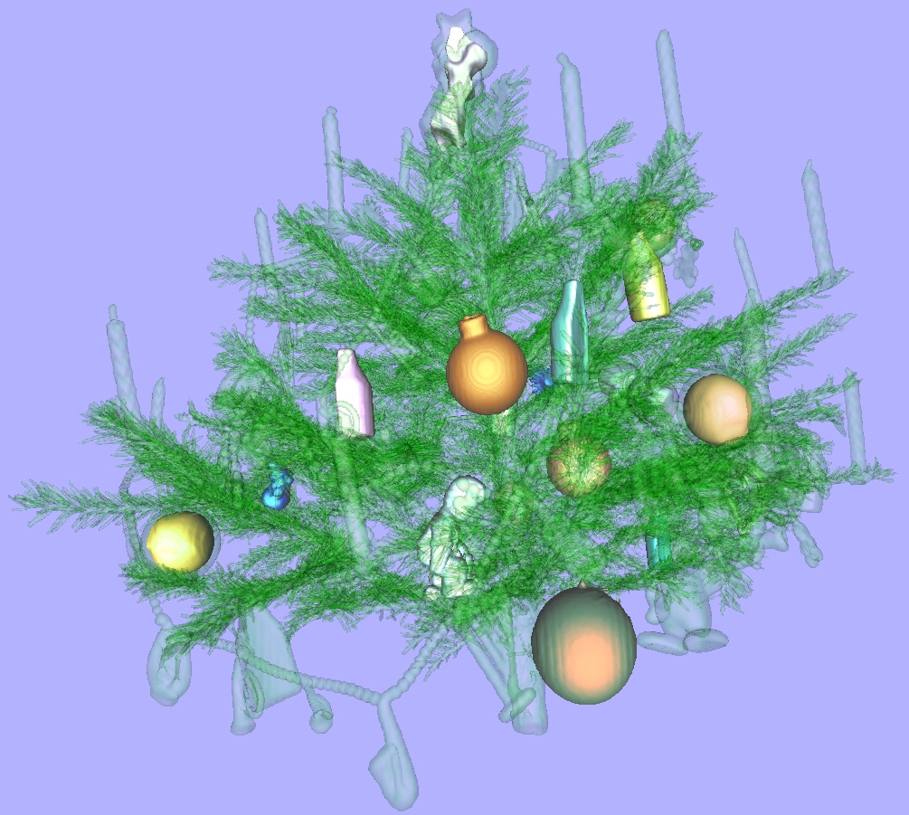
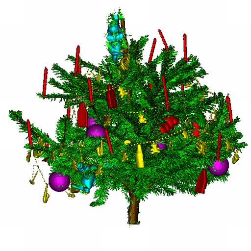
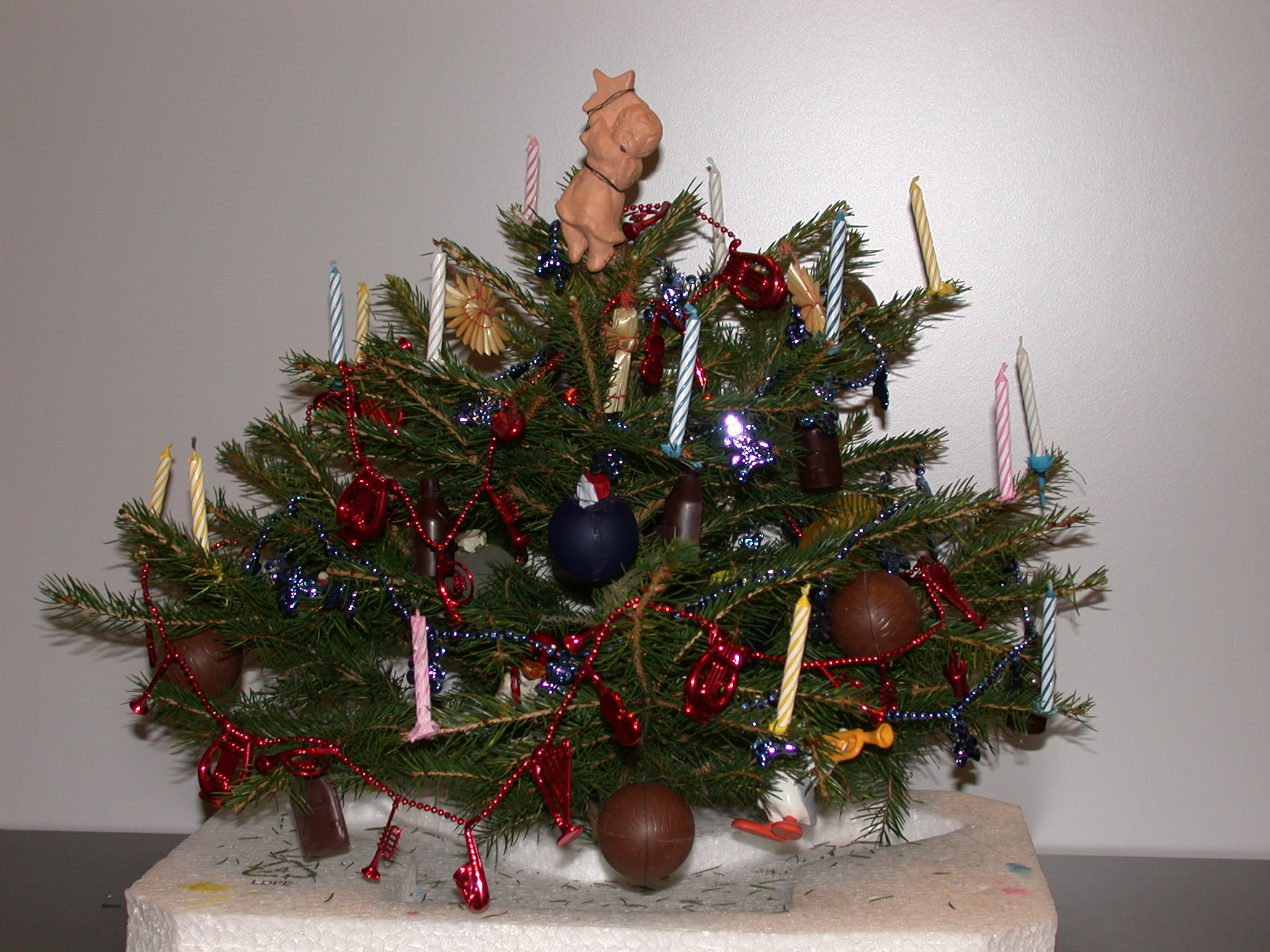
Model Preparation:Firstly, special care had to be taken in the preparation of the real model to assume a high data set quality. Especially, no metal was allowed to be used, as this would result in artifacts. Therefore the sweets were unwrapped and metal hooks replaced. Due to the limited diameter of the CT gantry, the size of the tree must not exceed 50 cm. Furthermore the proportions of the Christmas tree decoration and the small conifer should match those of a real Christmas tree. Therefore special items, as small candles for birthday cakes, were used.
|
|
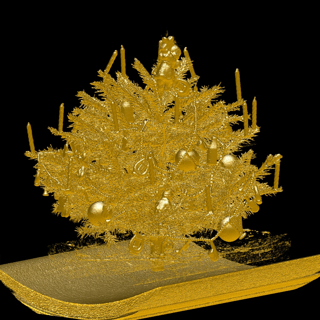
The CT acquisition protocoll had to be specifically designed as neither presets nor protocols are available for small trees. The Christmas-tree model was scanned with a Siemens Somatom Plus 4 Volume Zoom Multislice-CT scanner at the general hospital in Vienna. A tube voltage of 140kV and a tube current of 100mAs was used during the 40s scan. The collimation was 4 x 1mm, the table increment was 3mm/360� gantry rotation. Transverse sections with a nominal slice width of 1.25mm were reconstructed at 0.5mm intervals using a medium/sharp (B40) reconstruction kernel. A dataset of 512 x 512 x 998 voxels was reconstructed from a field of view of 476mm x 476mm x 499mm.
Download paper and slides
|
|
|
In proceedings of Visualization 2002, pp. 489-492
Best Case Study Award!
Boston, USA
Download data
Data format:Header:
2 byte - uSizeX
2 byte - uSizeY
2 byte - uSizeZ
Data: (16 bit - stored in unsigned short)
uSizeX * uSizeY * uSizeZ * sizeof(unsigned short)
- dataset-christmastree-128x124x128.zip
- dataset-christmastree-170x166x170.zip
- dataset-christmastree-256x249x256.zip
- dataset-christmastree-512x499x512.zip
Movie Gallery
|
|

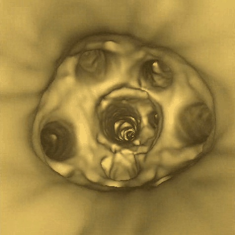
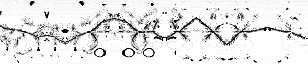
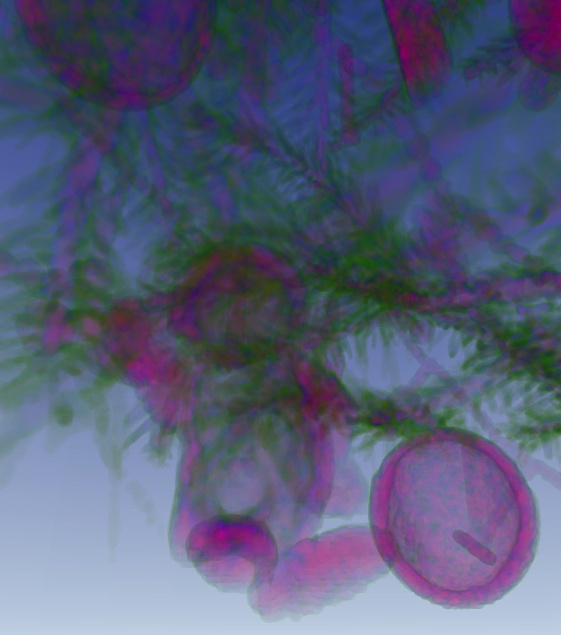
Image Gallery
|
|
|
|
|
|
|
|
|
|
|
|
|
|
|
|
|
|
|
|