

Abstract
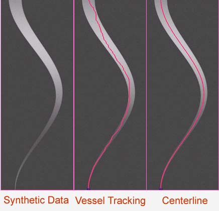
Purpose: The accurate determination of the central vessel axis is a prerequisite for automated visualization (curved planar reformation) and quantitation. The purpose of this work was to assess the accuracy of different algorithms for automated centerline detection in a phantom simulating the peripheral arterial tree.Methods and Material: Six algorithms were used to determine the centerline of a synthetic peripheral arterial vessel (aorto-to-pedal arteries, diameter 18-0.6mm) dataset (256x256x600, voxel size 0.5x0.5x0.5mm). They are ray-casting/thresholding (RCT), ray-casting/maximum gradient (RCMG), block matching (BM), fitting to ellipse (FE), center of gravity (CoG), and Randomized Hough transform (RHT). Gaussian noise whith a sigma: 0, 5 and 10 was used to observe the accuracy of the method under noise influence The accuracy of automatic centerline determination was quantified by measuring the error-distance between the derived centerlines, and the known centerline course of the synthetic dataset.
Results: BM demonstrated unacceptable performance in large vessels (>5mm) when the shift used was less than 3 voxels. RCMG demonstrated a greater error (mean of the error 4.73mm) in large diameter (>15mm) vessels than in small diameter (<15mm) vessels (mean of the error 0.64mm). Because RHT and FE use Canny edge detector preprocessing, both are sensitive to noise. CoG and RCT keep the mean of the error-distance significantly smaller (0.7mm and 0.9mm respectively) than all other algorithms.
Conclusion: CoG and RCT algorithms provide the most efficient centerline approximation over a wide range of vessel diameters.
Keywords:Centerline Detection, Vessel Segmentation, Medical Visualization